Conjoint Tendon Shoulder Anatomy / Medivisuals The Anatomy Of The Inguinal Area Medical Illustration : Unlike the other muscles in the anterior compartment of the arm, the biceps muscle crosses two joints, the shoulder joint and the elbow joint.
byAdmin•
0
Conjoint Tendon Shoulder Anatomy / Medivisuals The Anatomy Of The Inguinal Area Medical Illustration : Unlike the other muscles in the anterior compartment of the arm, the biceps muscle crosses two joints, the shoulder joint and the elbow joint.. Mar 27, 2020 · anatomy of the male pelvis on mr imaging: Muscle and tendon anatomy of the hip (adductors, gluteal muscles (or buttocks), hamstring muscles, femoral muscle quadrices). The different bursae of the hip region (trochanteric, ischial and iliopectineal bursae) The fascia on the lateral side of the conjoint tendon is incised to reveal the subscapularis external rotation puts the subscapularis fibers on stretch He used the arm to catch himself from falling.
Open and arthroscopic anterior shoulder stabilization. May 31, 2021 · internal abdominal oblique (musculus obliquus internus abdominis) internal abdominal oblique is a broad thin muscular sheet found on the lateral side of the abdomen.going from superficial to deep, the external abdominal oblique, internal abdominal oblique and transversus abdominis comprise the three distinct layers of the lateral abdominal wall. It forms the medial part of the posterior wall of the inguinal canal. Retraction of the conjoint tendon must be done with care. epub ahead of print fabricant pd, taylor sa, mccarthy mm, gausden e, moran cj, kang rw, cordasco fa.
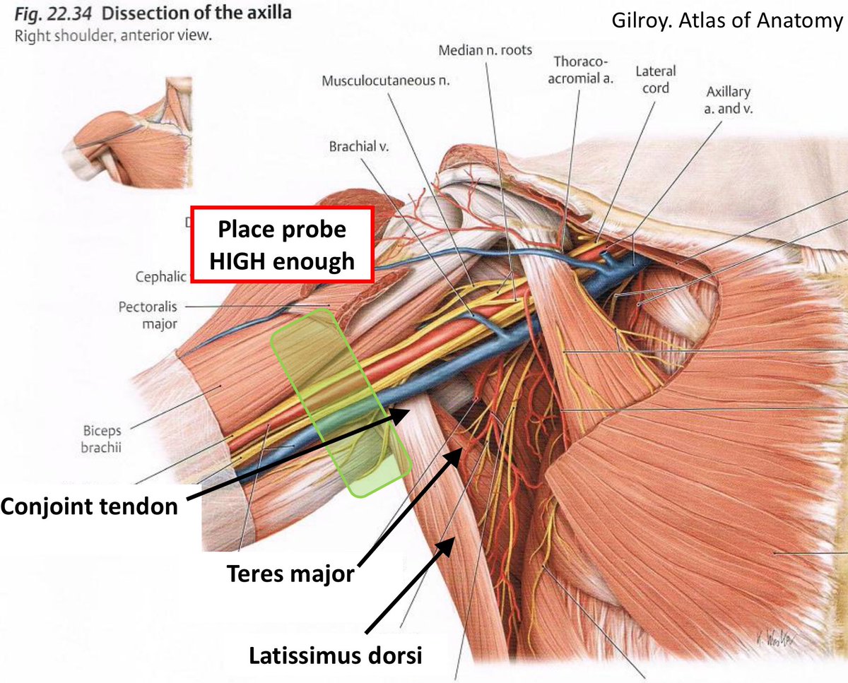
Ki Jinn Chin On Twitter These Images Are What I Base The Term Conjoint Tendon On Although I Appreciate The Structure Seen May Not Actually Arise From Teres Major Or Lat Dorsi from pbs.twimg.com The different bursae of the hip region (trochanteric, ischial and iliopectineal bursae) Sep 21, 2020 · muscles: He used the arm to catch himself from falling. He has not been wearing his sling and reports that he developed increased pain after slipping in the shower. It forms the medial part of the posterior wall of the inguinal canal. Retraction of the conjoint tendon must be done with care. On examination, he can flex the shoulder to 70 degrees, limited by pain. The fascia on the lateral side of the conjoint tendon is incised to reveal the subscapularis external rotation puts the subscapularis fibers on stretch
The different bursae of the hip region (trochanteric, ischial and iliopectineal bursae)
The teres minor (latin teres meaning 'rounded') is a narrow, elongated muscle of the rotator cuff.the muscle originates from the lateral border and adjacent posterior surface of the corresponding right or left scapula and inserts at both the greater tubercle of the humerus and the posterior surface of the joint capsule. Bursae of the lower limb: Incise the clavipectoral fascia lateral to the conjoined tendon and inferior the coracoacromial ligament. Open and arthroscopic anterior shoulder stabilization. On examination, he can flex the shoulder to 70 degrees, limited by pain. He has not been wearing his sling and reports that he developed increased pain after slipping in the shower. The conjoint tendon (previously known as the inguinal aponeurotic falx) is a sheath of connective tissue formed from the lower part of the common aponeurosis of the abdominal internal oblique muscle and the transversus abdominis muscle, joining the muscle to the pelvis. May 31, 2021 · internal abdominal oblique (musculus obliquus internus abdominis) internal abdominal oblique is a broad thin muscular sheet found on the lateral side of the abdomen.going from superficial to deep, the external abdominal oblique, internal abdominal oblique and transversus abdominis comprise the three distinct layers of the lateral abdominal wall. The fascia on the lateral side of the conjoint tendon is incised to reveal the subscapularis external rotation puts the subscapularis fibers on stretch Retract the deltoid muscle laterally using a delta (modified blunt hohmann) retractor and the conjoint tendon medially using a right angle retractor. The anatomy of the fascia lata and iliotibial tract; Unlike the other muscles in the anterior compartment of the arm, the biceps muscle crosses two joints, the shoulder joint and the elbow joint. It forms the medial part of the posterior wall of the inguinal canal.
epub ahead of print fabricant pd, taylor sa, mccarthy mm, gausden e, moran cj, kang rw, cordasco fa. Muscle and tendon anatomy of the hip (adductors, gluteal muscles (or buttocks), hamstring muscles, femoral muscle quadrices). He used the arm to catch himself from falling. The teres minor (latin teres meaning 'rounded') is a narrow, elongated muscle of the rotator cuff.the muscle originates from the lateral border and adjacent posterior surface of the corresponding right or left scapula and inserts at both the greater tubercle of the humerus and the posterior surface of the joint capsule. Extending from its origin on the coracoid, the tendon of the short head runs adjacent to the tendon of the coracobrachialis as the conjoint tendon.
Figure 33 26 From Proximal Humerus Resection The Tikhoff Linberg Procedure And Its Modifications Semantic Scholar from d3i71xaburhd42.cloudfront.net Identify the coracoid process and the conjoined tendon. epub ahead of print fabricant pd, taylor sa, mccarthy mm, gausden e, moran cj, kang rw, cordasco fa. Retract the deltoid muscle laterally using a delta (modified blunt hohmann) retractor and the conjoint tendon medially using a right angle retractor. The different bursae of the hip region (trochanteric, ischial and iliopectineal bursae) Retraction of the conjoint tendon must be done with care. The conjoint tendon (previously known as the inguinal aponeurotic falx) is a sheath of connective tissue formed from the lower part of the common aponeurosis of the abdominal internal oblique muscle and the transversus abdominis muscle, joining the muscle to the pelvis. May 31, 2021 · internal abdominal oblique (musculus obliquus internus abdominis) internal abdominal oblique is a broad thin muscular sheet found on the lateral side of the abdomen.going from superficial to deep, the external abdominal oblique, internal abdominal oblique and transversus abdominis comprise the three distinct layers of the lateral abdominal wall. Midterm clinical outcomes for arthroscopic subdeltoid transfer of the long head of the biceps tendon to the conjoint tendon.
On examination, he can flex the shoulder to 70 degrees, limited by pain.
epub ahead of print fabricant pd, taylor sa, mccarthy mm, gausden e, moran cj, kang rw, cordasco fa. The teres minor (latin teres meaning 'rounded') is a narrow, elongated muscle of the rotator cuff.the muscle originates from the lateral border and adjacent posterior surface of the corresponding right or left scapula and inserts at both the greater tubercle of the humerus and the posterior surface of the joint capsule. The anatomy of the fascia lata and iliotibial tract; The fascia on the lateral side of the conjoint tendon is incised to reveal the subscapularis external rotation puts the subscapularis fibers on stretch Mar 27, 2020 · anatomy of the male pelvis on mr imaging: The different bursae of the hip region (trochanteric, ischial and iliopectineal bursae) Incise the clavipectoral fascia lateral to the conjoined tendon and inferior the coracoacromial ligament. He has not been wearing his sling and reports that he developed increased pain after slipping in the shower. Bursae of the lower limb: Retract the deltoid muscle laterally using a delta (modified blunt hohmann) retractor and the conjoint tendon medially using a right angle retractor. Extending from its origin on the coracoid, the tendon of the short head runs adjacent to the tendon of the coracobrachialis as the conjoint tendon. May 31, 2021 · internal abdominal oblique (musculus obliquus internus abdominis) internal abdominal oblique is a broad thin muscular sheet found on the lateral side of the abdomen.going from superficial to deep, the external abdominal oblique, internal abdominal oblique and transversus abdominis comprise the three distinct layers of the lateral abdominal wall. Open and arthroscopic anterior shoulder stabilization.
Incise the clavipectoral fascia lateral to the conjoined tendon and inferior the coracoacromial ligament. Unlike the other muscles in the anterior compartment of the arm, the biceps muscle crosses two joints, the shoulder joint and the elbow joint. He used the arm to catch himself from falling. Identify the coracoid process and the conjoined tendon. Mar 27, 2020 · anatomy of the male pelvis on mr imaging:
Y1s2 Gi Block Anatomy Inguinal Region Flashcards Quizlet from o.quizlet.com Retraction of the conjoint tendon must be done with care. epub ahead of print fabricant pd, taylor sa, mccarthy mm, gausden e, moran cj, kang rw, cordasco fa. Retract the deltoid muscle laterally using a delta (modified blunt hohmann) retractor and the conjoint tendon medially using a right angle retractor. Incise the clavipectoral fascia lateral to the conjoined tendon and inferior the coracoacromial ligament. He used the arm to catch himself from falling. May 31, 2021 · internal abdominal oblique (musculus obliquus internus abdominis) internal abdominal oblique is a broad thin muscular sheet found on the lateral side of the abdomen.going from superficial to deep, the external abdominal oblique, internal abdominal oblique and transversus abdominis comprise the three distinct layers of the lateral abdominal wall. He has not been wearing his sling and reports that he developed increased pain after slipping in the shower. It forms the medial part of the posterior wall of the inguinal canal.
epub ahead of print fabricant pd, taylor sa, mccarthy mm, gausden e, moran cj, kang rw, cordasco fa.
Open and arthroscopic anterior shoulder stabilization. Retract the deltoid muscle laterally using a delta (modified blunt hohmann) retractor and the conjoint tendon medially using a right angle retractor. Sep 21, 2020 · muscles: May 31, 2021 · internal abdominal oblique (musculus obliquus internus abdominis) internal abdominal oblique is a broad thin muscular sheet found on the lateral side of the abdomen.going from superficial to deep, the external abdominal oblique, internal abdominal oblique and transversus abdominis comprise the three distinct layers of the lateral abdominal wall. Incise the clavipectoral fascia lateral to the conjoined tendon and inferior the coracoacromial ligament. The anatomy of the fascia lata and iliotibial tract; Mar 27, 2020 · anatomy of the male pelvis on mr imaging: Unlike the other muscles in the anterior compartment of the arm, the biceps muscle crosses two joints, the shoulder joint and the elbow joint. Identify the coracoid process and the conjoined tendon. The different bursae of the hip region (trochanteric, ischial and iliopectineal bursae) He has not been wearing his sling and reports that he developed increased pain after slipping in the shower. The conjoint tendon (previously known as the inguinal aponeurotic falx) is a sheath of connective tissue formed from the lower part of the common aponeurosis of the abdominal internal oblique muscle and the transversus abdominis muscle, joining the muscle to the pelvis. Bursae of the lower limb: